Before & after
Real results. Real consent.
A clinically respectful gallery of authentic patient photographs, shown only with documented permission.
No cases published yet.
We will not fill this page with stand-in, retouched or AI-generated images. When patients have given written permission for public display, their cases will appear here with the procedure, relevant details and consistent framing.
Individual results vary. Photographs show what was possible for one person, not what to expect for you.
Drag, or use the arrow keys, to compare. This is how real cases will work.
Educational diagrams
How breast anatomy and surgery relate
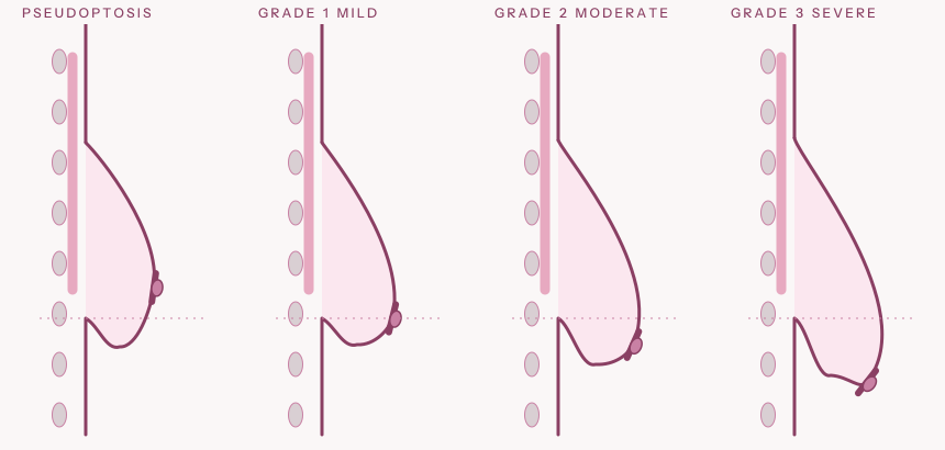
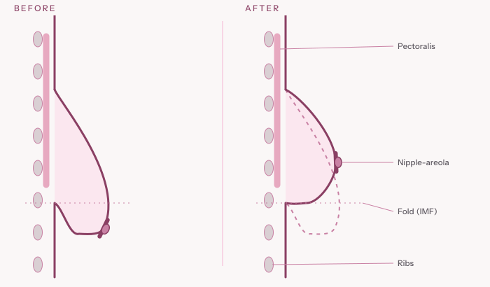
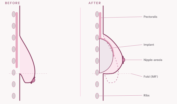
Side-view teaching diagrams showing the ribs, pectoralis major muscle, skin envelope, inframammary fold (IMF) and nipple position. They are schematic drawings, not photographs and not patient results. Dashed line: the starting contour.
Grading breast ptosis (sagging)
What each operation aims to change
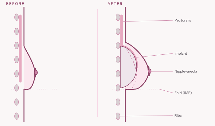
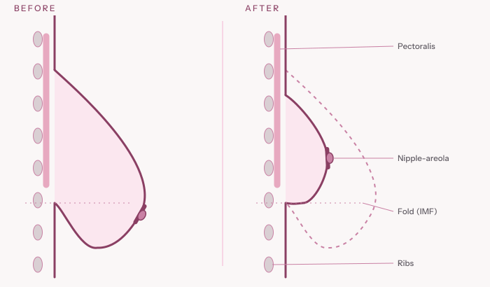
Where an implant sits
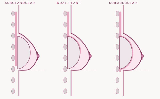
Educational illustration only. Individual outcomes vary and depend on anatomy, technique and healing. Your surgeon will explain what is realistic for you.
Our standards
What you will see.
- Only authentic photographs of real patients, with documented consent for this use.
- Consistent lighting, angle and framing, with no filters or body-altering edits.
- The procedure and relevant case information where the patient has approved it.
- A clear note that individual results vary.
- Clinical photographs kept visually separate from brand illustrations, so art is never mistaken for outcomes.
Your questions.
Your goals.
Your consultation.
A consultation is a conversation: your goals, your anatomy, your options and the honest trade-offs. Booking one is not booking surgery.